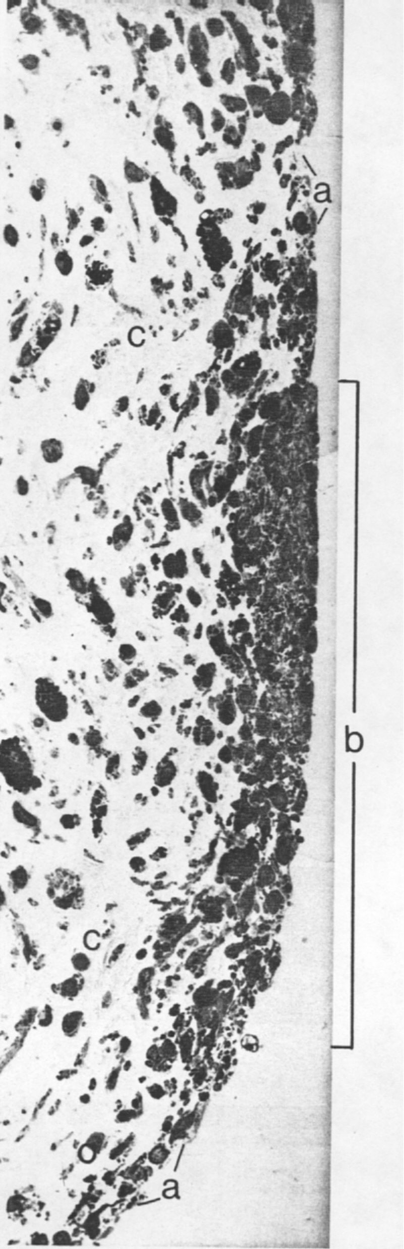
A pigmented iris showing the anterior border layer (a) and an iris freckle (b). In the area of the freckle the anterior border layer becomes more cellular. The iris stroma (c) beneath the freckle also is more cellular. (X 400.)
|
|
Figure I-13
A pigmented iris showing the anterior border layer (a) and an iris freckle (b). In the area of the freckle the anterior border layer becomes more cellular. The iris stroma (c) beneath the freckle also is more cellular. (X 400.) |
|||
|
|
||||