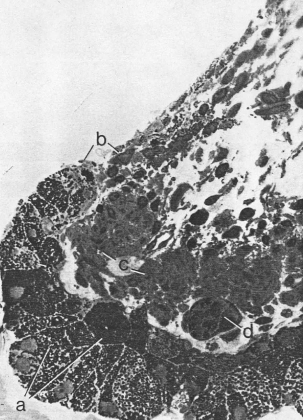
Pupillary margin of the iris to show the two pigment epithelial layers (a) and their continuity with the anterior border layer of the iris (b). The sphincter muscle of the iris (c) is slightly curved near the pupillary margin and is continuous with the anterior border layer by a thin layer of connective tissue. A clump cell measuring 90 microns (d) is seen near the sphincter muscle. (X 680.)