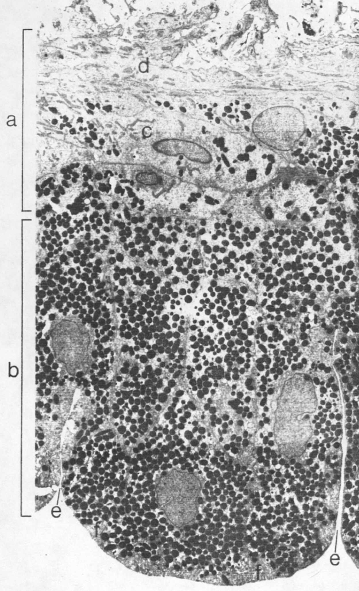
Low-power electron micrograph of the two layers of iris epithelium. The anterior epithelium and dilator muscle are shown at (a) and the posterior epithelium at (b). The epithelial portion of the anterior epithelium is at (c) and the muscular component at (d). Note the paucity of melanin granules in the anterior epithelium and their distribution in the apical portion near the nucleus. The posterior iris surface shows two deep furrows (e) and the basal epithelium shows extensive infolding (f). (X2500.)